Symptoms of cerebral facial angiomatosis?
summary
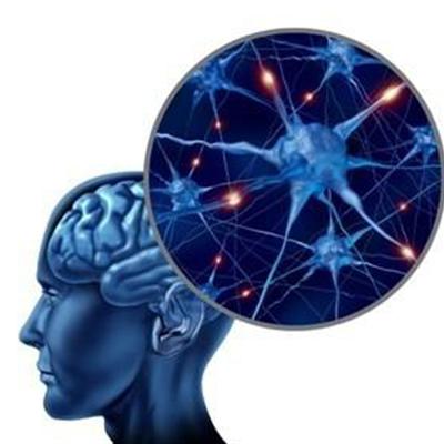
Facial hemangioma (equivalent to trigeminal nerve distribution area) and similar vascular carcinoma on the surface of corresponding cerebral hemisphere are called encephalofacial angiomatosis. It will affect our normal life. In 879 and 1929, it was described for Sturge and Weber, so it was named Sturge Weber disease. Is a rare facial hemangioma, contralateral limb twitch or hemiplegia, ipsilateral intracranial calcification and intracranial hemangioma disease, mental retardation as the main characteristics of the nervous system genetic disease, next I will introduce the symptoms of cerebral facial hemangioma disease?.
Symptoms of cerebral facial angiomatosis?
First: the edge of vascular nevus is clear, slightly higher than the skin, and it will not fade when pressed. Common seizures, intellectual decline, cerebral hemangioma on the opposite side of hemiplegia and hemiatrophy. Glaucoma and exophthalmos, congenital abnormalities, such as iris defect and lens opacity.
Second, angiography. DSA can find abnormalities of capillaries and veins, hyperplasia of capillaries on the surface of affected hemisphere, significant reduction of veins and hypoplasia of superior sagittal sinus
Third: look at EEG again. EEG showed low amplitude in the affected hemisphere, α The decrease of wave intensity was consistent with the degree of intracranial calcification; Epileptogenic waves can be seen.
matters needing attention
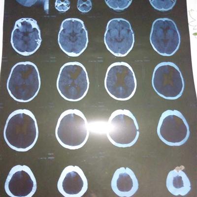
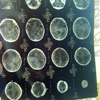
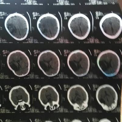
Patients usually accompanied with epilepsy, glaucoma, exophthalmos, contralateral hemiplegia, hemiatrophy and other symptoms. The double track calcification on X-ray, CT and MRI are helpful in the diagnosis of cerebral atrophy and meningeal hemangioma.