Got pharyngeal hind space infection how should do?
summary
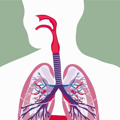
Retropharyngeal space infection is a suppurative infection that occurs between the pharynx, the posterior wall of the esophagus and the prevertebral fascia (Fig. 1). Most of them are caused by nasal and ear infection or cervical tuberculosis. Clinical early respiratory tract infection, gradually forming a posterior pharyngeal abscess. Got pharyngeal hind space infection how should do? Next, I'd like to share my views with you.
Got pharyngeal hind space infection how should do?
Transoral approach: the most commonly used, suitable for early respiratory obstruction or other complications and can use local anesthesia. Take the supine head down position (Fig. 2) to prevent the pus from flowing into the trachea. Under the condition of good lighting and suction, the tongue base was pressed on the bottom of the mouth with a tongue depressor to see the abscess clearly. Puncture and suction were carried out at the most protruding part. After the pus was extracted as much as possible, a 1-2cm vertical incision was made at the low position of the abscess (close to the laryngopharynx end) with a long handle knife (wrapped with adhesive tape or spinning strip to expose only 1cm tip), In order to avoid injury to the large blood vessels on the neck side), while suction, while incision. Then the curved vascular clamp was inserted into the abscess cavity to enlarge the incision, and the pus was drained and sucked out without drainage.
Anterolateral cervical approach (Dean operation): it is suitable for patients with large or low retropharyngeal space abscess, retropharyngeal space cellulitis, parapharyngeal space infection, mediastinitis, sepsis and other complications. Under local or general anesthesia, the patient was supine and the head was inclined to the contralateral side. A transverse incision was made at the front edge of sternocleidomastoid muscle, between hyoid bone and sternum. The sternocleidomastoid muscle and carotid sheath were pulled to the outside, and the thyroid gland, superior thyroid vessels and superior laryngeal nerve were pulled to the inside. The abscess is usually exposed in the hypopharyngeal plane. For good exposure, the middle thyroid vein, inferior thyroid artery and scapulohyoid muscle can be cut off, and the hypoglossal nerve, lingual blood vessel and facial blood vessel can be reserved. After aspiration, the abscess was opened between the carotid sheath and the hypopharyngeal constrictor muscle (the abscess was punctured with closed blunt vascular forceps, and then the forceps were opened to enlarge the drainage).
Lateral cervical approach: the indications are the same as anterolateral cervical approach. Under local or general anesthesia, the patient lies on his back with his head tilted to the contralateral side, so that the neurovascular bundle is pulled away from the spine. The skin incision was made along the posterior edge of sternocleidomastoid muscle to avoid the injury of neck vessels and nerves.
matters needing attention
The etiology of this disease is mainly due to infection, usually following the development of oral periodontal and gingival, tonsillar infection caused by the spread, so actively treat the maxillary third molar pericoronitis, periapical periodontitis and tonsillitis, posterior alveolar nerve block anesthesia, foramen ovale anesthesia, temporal trigeminal sympathetic nerve block to prevent infection. At the same time, we should take active anti infection treatment for the infected cases to avoid the occurrence of severe complications such as encephalitis and sepsis.