What is the manifestation of enlarged mediastinal voiced boundary?
summary
The enlargement of mediastinal voiced boundary was mainly based on clinical manifestations, such as penetrating chest trauma, rupture of esophagus or trachea, perforation of esophagus caused by foreign body in pharynx, anastomotic leakage after esophageal surgery, perforation of trauma during esophagoscopy and perforation of esophageal cancer ulcer. It can be used for imaging examination. What is the manifestation of enlarged mediastinal voiced boundary?
What is the manifestation of enlarged mediastinal voiced boundary?
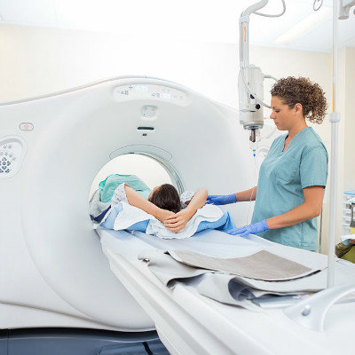
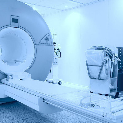
Diagnosis is mainly based on clinical manifestations, but because mediastinitis is a part of the whole process of infection, and simple mediastinitis in X-ray chest film, in addition to the possibility of mediastinal shadow widened, mediastinal emphysema, there is no special performance, so clinical see more is limited mediastinal abscess, or posterior mediastinum in the lateral chest film has gas level, emphysema and other manifestations, sometimes the diagnosis is not easy. In X-ray examination, anteroposterior and lateral chest films are very important. General bedside photography is not clear due to the relationship between projection conditions. For the convenience of diagnosis, it is better to take the chest film of half sitting position and lateral position. If the rupture of esophagus or trachea is suspected, 40% sterile lipiodol can be used for radiography, and barium should be avoided to avoid long-term retention and stimulate the tissue.
The thoracic cavity of the human body is divided into two pleural cavities, the middle part of which is called mediastinum. Mediastinum contains heart, thoracic blood vessels, trachea, esophagus, nerve and lymphoid tissue. The mediastinum can be divided into several regions. From the sternal angle (that is, the junction of sternal stalk and sternal body, which can be felt on the body surface as an obvious transverse ridge), a horizontal line is drawn backward to the lower edge of the fourth thoracic vertebral body. Above this line is called the upper mediastinum, and below this line is called the lower mediastinum. The upper mediastinum is bounded by trachea. The anterior part is the anterior upper mediastinum, and the posterior part is the posterior upper mediastinum. The lower mediastinum is divided into three parts: anterior, middle and posterior. The anterior part of the pericardium is the anterior mediastinum, the middle mediastinum is where the pericardium is, and the posterior mediastinum is between the pericardium and the spine.
The anterior superior mediastinum is mainly composed of thymus and intrathoracic thyroid. The posterior superior mediastinum is composed of trachea, esophagus, aortic arch and its three brachiocephalic vascular branches, thoracic duct, vagus nerve, etc. The lower part of thymus, lymph nodes, fat and connective tissue were found in the lower anterior mediastinum. There are esophagus, thoracic duct, descending aorta and its branches, azygous vein, hemiazygous vein, vagus nerve and sympathetic nerve in the lower posterior mediastinum.
matters needing attention
For acute mediastinitis, the first thing is to treat the etiology, control infection and support therapy (blood transfusion, infusion, oxygen). When chronic mediastinitis is accompanied by severe superior vena cava obstruction, surgery is needed to establish collateral circulation and bypass. The treatment of mediastinal hernia, mainly the treatment of primary disease, remove the cause of disease, can make the mediastinal hernia recover quickly. Mediastinal emphysema, if only a small amount of gas, can not be treated by itself. Severe cases are also the causes of treatment (such as trauma, emphysema, rupture of pulmonary bullae, etc.). If the slow absorption of gas causes dyspnea or affects the pronunciation of the patient, an incision on the sternal notch can be made to exhaust the subdermal tissue.