Hydatid disease symptoms?
summary
Echinococcosis can occur in the human body for several to several decades. The clinical manifestations vary with the parasitic site, cyst size and complications. Due to the different species of parasites, the clinical manifestations are cystic echinococcosis (unicameral echinococcosis), alveolar echinococcosis (multilocular echinococcosis) and mixed echinococcosis. Multilocular echinococcosis is mostly caused by the larvae of Echinococcus vonii or Echinococcus oligoarthrum. It is found in central and South America abroad, but has not been found in China. Hydatid disease symptoms? Let's talk about it
Hydatid disease symptoms?
The right upper abdomen mass appears when the cystic pericarditis is extremely enlarged. The patient has a sense of fullness and may have compression symptoms. Most of the cysts were located in the right lobe, and most of them were located on the surface. When the cyst is located in the center of the right lobe, the liver is diffusely enlarged and develops upward, which can cause reactive pleural effusion and atelectasis; If it develops downward and forward, it will bulge into abdominal cavity. Most of the patients found that the liver was extremely swollen, local surface smooth cyst. A few cases can hear tremor after percussion on cyst. Most of the liver functions were normal and the ratio of albumin to globulin was inverted. B-mode ultrasound, radioisotope scan and CT scan all showed space occupying lesions. Echinococcosis caused by Echinococcus granulosus is usually called unilocular echinococcosis; It is called multilocular echinococcosis, which is caused by echinococcosis. Echinococcosis proliferates in an invasive manner, similar to malignant tumor. Hepatic alveolar echinococcosis can also be metastasized through lymph or blood, secondary to pulmonary and cerebral alveolar echinococcosis, so it is called malignant echinococcosis. The liver became hard and uneven.

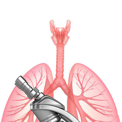
The lung tissue is relatively loose, so the hydatid sac grows fast, often with dry cough, hemoptysis and other symptoms. About 2 / 3 of the patients' lesions were located in the right lung, and most of them were in the lower lobe. In the cases without complications, X-ray examination showed single or multiple round, oval or multiple ring masses with clear and smooth edges (fuzzy edges in case of secondary infection). Cysts deform with respiration, rarely calcify, and vary in size. The cyst was punctured and the cyst fluid was completely discharged, showing cavity type on X-ray; Severe hydropneumothorax may occur when the cyst breaks into the chest. About half of the patients' cysts broke into the bronchus and healed with the cough up of the cystic fluid. Occasionally, a large amount of cystic fluid overflowed and caused asphyxia.

The incidence rate is low (1% to 2%), most commonly seen in children, with the most common parietal lobe. The clinical manifestations are seizures and intracranial hypertension. Most of the cysts are single, most of them are located under the cortex. In cases with extensive lesions, the lateral ventricle can be involved, and the skull can be compressed and eroded. Cerebral angiography, CT and MRI are helpful in diagnosis.

matters needing attention
1. Strengthen and popularize health education. 2. Registration management of domestic dogs and strict control of ownerless dogs. 3. Treat sick dogs. 4. Strictly manage market and family slaughtering to prevent dogs from contacting organs infected by hydatid.