Early symptoms of heat shock erythema?
summary
Erythema ab igne, also known as heat shock erythema, is a kind of persistent telangiectasia reticular erythema caused by repeated exposure of local skin to high temperature stimulation that is not enough to cause burns. Sometimes the pigment increases significantly, which is called fire shock reticular pigmentation. This disease is similar to "huolongchuang" recorded in the literature of traditional Chinese medicine. For example. What's the secret of surgery? Huohuang sore records: "to avoid cold fire, long burning skin, fire gas into the sore, sweating pain." Let's talk about the early symptoms of heat shock erythema.
Early symptoms of heat shock erythema?
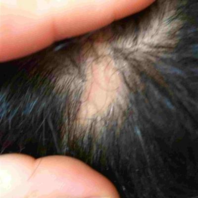
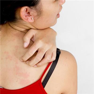
It is mainly seen in those who are engaged in high temperature operation or who are weak and warm for a long time. After several hours of local skin exposure to hot environment, it showed flushing or temporary reticular erythema (caused by telangiectasia and congestion). If repeatedly exposed to heat source, the erythema is obvious, showing purplish red or purplish brown reticular erythema (caused by venous dilatation) and accompanied by pigmentation, which often occurs after cure.

For persistent pigmentation, decolorizing agents such as 3% ~ 5% hydroquinone cream or superoxide dismutase (SOD) cream and 0.05% ~ 0.1% tretinoin ointment can be used. Atrophic lesions are mostly permanent. Hyperkeratosis can be removed with 5-FU ointment or surgery. Traditional Chinese medicine can use Qingliang ointment or Shigella oil. Can also spread Qushi powder.

It will last forever. Occasionally, blisters, mild skin atrophy and hyperkeratosis may occur. The distribution of skin lesions was consistent with the area of heat and radiation, such as sleeping on hot Kang and electric blanket, which mostly occurred on the back; People who sit for warmth are mostly at the extension side of the lower leg. I felt burning and itching.

matters needing attention
1. Reticular lesions were dilated capillaries without pigmentation and scales, which were symmetrical and diffuse. 2. The telangiectasia and atrophy of atrophic skin heterochromatosis are more prominent.