How much does a renal arteriography cost?
summary
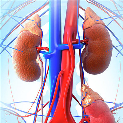
The cost of renal arteriography ranges from 3000 to 4000 yuan. The cost varies according to the contrast medium used. This examination is mainly used in two aspects. The first case is renal vascular bleeding. Under the contrast medium, the bleeding small vessels can be found for interventional embolization. In the second case, when the renal artery stenosis occurs, the location and degree of the stenosis can be determined by angiography. According to the clinical manifestations and renal function, stent placement can be selected.
How much does a renal arteriography cost?
Angiography steps: abdominal aortography should be performed first to observe the blood supply of bilateral renal arteries, distinguish whether there are accessory renal arteries and aberrant renal arteries, evaluate the renal function of both sides, and then perform the renal artery and target angiography of the affected side to determine the target vessels of bleeding or lesions. When embolizing, we should try our best to super select intubation, use embolic agent properly, control the total amount of embolization, avoid reflux and protect normal renal tissue. In case of arteriovenous malformation or fistula, it is necessary to embolize the pulmonary artery with spring coil or memory conch coil.

The catheter should be withdrawn from the target vessel and located in the main renal artery to avoid high-speed contrast agent impact and embolism. At the same time, combined with small renal vein branch injury, embolizing the injured artery branch and hemostasis treatment can often be effective. If there is still active bleeding after interventional treatment, a catheter can be placed in the renal artery, and pituitrin can be perfused at 0.2u/min for 8-10h. The color of urine, red and white blood, hematocrit and other indicators can be observed, and the perfusion dose can be adjusted.
Superselective angiography of renal artery: superselective angiography of target vessel was performed by changing catheter to determine the location, scope and vascular anatomy of bleeding artery and formulate treatment plan. The angiographic findings of acute renal hemorrhage were as follows: the renal branches were interrupted or absent, and the branches were reduced; Irregular peripheral blood vessels, extravasation of contrast media or traumatic pseudoaneurysmal changes were found; The laceration area showed irregular strip defect or renal parenchyma separation; In severe contusion and laceration, the size and shape of the renal tissue cleft or the renal pole blood vessels connected with the large blood vessels could be shown in its substantive phase, and its edge was rough and irregular; The density of renal pole laceration area decreased or did not develop; In the parenchymal phase, the contour of kidney was deformed; Arteriovenous fistula; Subcapsular hematoma displaces or dilates the encapsulated artery.

matters needing attention
Renal artery embolization: superselective catheterization of target vessels. Gelatin sponge particles or PVA particles were dissolved in normal saline, and an appropriate amount of contrast agent was added. Under TV monitoring, the embolic agent was slowly injected according to the volume of embolic vessels. The disappearance of arterial branches in the lesion area and the retention of contrast medium in the vessel or injured area for a long time indicate the success of embolization.