Methods of esophageal cancer examination
summary
During this period of time, my grandmother felt burning pain in her sternum after eating, and it was a little difficult for her to swallow. She had to drink a lot of water when eating. In order to prevent this situation, I'd like to introduce the examination method of esophageal cancer.
Methods of esophageal cancer examination
First: the main disease is to carry out appropriate examination, because of the occurrence of esophageal cancer, there will be hope of treatment after examination. Endoscopic ultrasonography (EUS) and endoscopic ultrasonography (EUS) are gradually used in clinic. The advantages of this method are that it can accurately measure the depth of the lesion in the esophageal wall; it can measure the extramural abnormal enlarged lymph nodes; it can easily distinguish the lesion in the esophageal wall, which is a useful method for the diagnosis of esophageal cancer. Esophageal exfoliative cytology examination, this method is simple, less pain, low false positive rate, practice has proved that in the high incidence area of esophageal cancer for large-scale census is feasible, the total positive rate can reach more than 90%, is the preferred method of early diagnosis of esophageal cancer.

Second: X-ray barium meal examination, in the face of disease examination, in fact, there will be a lot of methods, you should also pay attention to the use of appropriate treatment measures in life. In addition to the difficulty of showing very early esophageal cancer, experienced radiologists fully adjusted barium to make the patient swallow in small mouthfuls in several times, carefully observed in multiple directions and air barium double contrast, most of them could find esophageal mucosa thickening, tortuosity or dotted line interruption, or hair on the edge of esophagus, or small filling defect, or small niche, or local tube wall stiffness, etc; It seems that this method can also be used in the diagnosis of esophageal cancer.

Third: chest CT scan, such inspection measures in our lives will be very common, but, in the face of such diseases, is also effective. There are different opinions about its role in the diagnosis of esophageal cancer, but it is helpful for the staging of esophageal cancer, the judgment of resection possibility and the estimation of prognosis. Fiberoptic endoscopy has gradually replaced metal rigid tube endoscopy. Because of its flexibility, good lighting, wide vision, safety and accuracy, it has become a reliable method for routine clinical diagnosis, postoperative follow-up and curative effect observation of upper gastrointestinal diseases (esophageal cancer, gastric cancer, etc.). In the early stage of esophageal cancer, the detection rate of fiber endoscopy can reach more than 85%.
matters needing attention
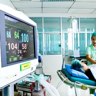
Postoperative nursing because of the special location of the disease, so patients after surgery try to reduce neck activity, neck do anastomosis patients after surgery also try to reduce neck activity, so that it can be relatively fixed, in order to facilitate the early healing of the anastomosis. If postoperative high fever, dyspnea and other adverse conditions, we must promptly inform the doctor.