How does right femur head epiphysis ischemia necrosis do?
summary
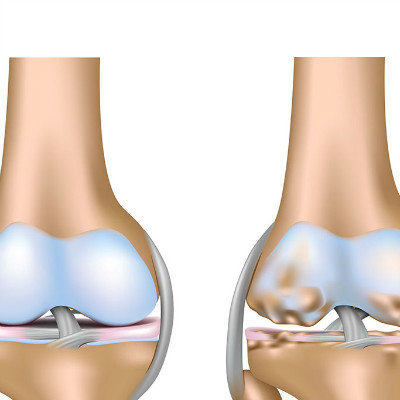
Avascular necrosis of the femoral head epiphysis, also known as Legg cave Perthes disease, osteochondritis of the femoral head and flat hip, is a common osteochondral necrosis. Most scholars believe that the main causes of the disease are the obstruction of internal venous drainage and the increase of intraosseous pressure. How does that right femur head epiphysis ischemia necrosis do?
How does right femur head epiphysis ischemia necrosis do?
Patients with femoral head necrosis should be prepared for a long time and have full confidence in the treatment. In terms of drugs, it is suggested that you should insist on taking some Chinese patent medicines, such as Tongluo Shenggu Capsule, to improve the blood circulation of the femoral head, promote the regeneration of new bone, avoid taking hormone drugs, and pay attention to protect the femoral head in life, avoid heavy load and trauma.
X-ray plain film is the main examination method, but it can not show the initial lesions. CT examination can show suspicious or difficult to show early repair changes of necrosis. MRL is a sensitive and specific method in the diagnosis of early necrosis of the disease. It can be used in the diagnosis of patients with negative plain film and CT. At the initial stage, there was no change in bone, only mild widening of the hip joint space, slight outward movement of the femoral head, swelling of the soft tissue above the joint capsule, distortion or blurring of the normal fat space, and the shape and size of the femoral head remained normal.
In the late stage, if the clinical treatment is timely, the necrotic bone of the epiphysis can be absorbed, fragmented and disappeared, and new bone gradually appears. The bone structure can be restored to normal, and the epiphysis can gradually return to its smooth and neat shape. If the treatment is delayed or improper, the femoral head is often left with fungiform or round cap shape deformity, the femoral neck is short and thick, the greater trochanter is elevated, the head is inclined forward and downward, the neck shaft angle is smaller, resulting in coxa vara and hip subluxation, and the upper part of acetabulum is straight and irregular; In some patients, the hip joint space became narrower and secondary degenerative osteoarthropathy appeared.
matters needing attention
This disease is mainly differentiated from tuberculosis of the hip joint. The latter is characterized by limited bone destruction of the epiphysis of the femoral head, progressive aggravation, even complete disappearance of the epiphysis, less bone sclerosis around the bone destruction, extensive osteoporosis of the adjacent joint, earlier joint space narrowing, no obvious epiphyseal plate and metaphyseal widening.