Choriocarcinoma symptoms?
summary
Gynecological disease is a malignant tumor originated from embryonic chorion. It is a malignant tumor originated from embryonic chorion, including all of the two trophoblasts. It often occurs in the uterus, but it is not the only primary site. About 50% of choriocarcinoma occurred after hydatidiform mole.
Choriocarcinoma symptoms?
22% occurred after normal delivery; Others occurred during ectopic pregnancy. This is a rare malignant tumor, mainly in women under 35 years old. The common clinical symptoms of choriocarcinoma are continuous irregular vaginal bleeding, the amount of blood is uncertain, sometimes the first temporary amenorrhea, and then sudden vaginal bleeding.

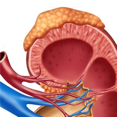
Examination can find that the uterus is enlarged and soft, irregular shape. The vagina has red and stinky bloody secretions. The general symptoms are anemia, emaciation, and even cachexia, which may be accompanied by fever. Because of the high degree of malignancy of the disease, early lung metastasis, chest pain, cough, hemoptysis and other symptoms can occur. The other metastasis sites were vagina, vulva, pelvic cavity, liver, brain and so on.

The main basis for the diagnosis of this disease is: after postpartum or abortion, especially after hydatidiform mole, vaginal bleeding continues, uterine involution is poor, the uterine body is large and soft, urine pregnancy test continues to be positive, the symptoms of curettage are not improved, and the chest X-ray film shows nodular lung. It should be considered as the disease because of the cotton ball or flake shadow. The determination of human chorionic gonadotropin (hCG) has important reference value in the diagnosis of this disease. Choriocarcinoma can be diagnosed by curettage, but sometimes the tumor is located between the muscle walls.

matters needing attention
Histologically, choriocarcinoma is very different from other cancers. Choriocarcinoma has no inherent connective tissue stromal cells, only necrotic foci composed of trophoblasts, blood clots and coagulative necrotic tissues, and no inherent blood vessels. Cancer cells directly contact with the host blood for nutrition. In the center of the tumor, cancer cells are often not found. The closer to the edge of the tumor, the more obvious the tumor cells are. However, villi can not be seen, only clusters of trophoblast can be seen.