How is trabecular meshwork pigmentation to return a responsibility?
summary
Trabecular meshwork pigmentation is the clinical diagnosis of pigmented glaucoma. Pigmented glaucoma is a secondary open-angle glaucoma caused by anterior segment pigment dissemination. Usually, it is easy to be confused with iris depigmentation and uveitis symptoms. Generally, drug treatment is used first, followed by laser treatment, and finally surgical treatment is considered.
How is trabecular meshwork pigmentation to return a responsibility?
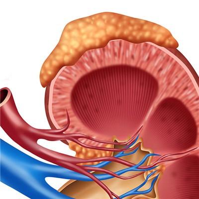
The main basis is the signs of PDS and the changes of intraocular pressure, glaucomatous visual field and optic disc. The most characteristic part is the radial light transmission area of the iris in the middle part. Another slit lamp examination showed Krukenberg fusiform pigmentation in the posterior wall of cornea, deep anterior chamber, retroversion of iris with pigmentation, and pigmentation near the equator on the posterior surface of lens after pupil dilation. Iridocorneal keratoscopy showed wide angle and dense pigmentation on trabecula. With iris signs and some other signs, PDS can be diagnosed; PG can be diagnosed if it is accompanied by the changes of visual field and optic disc in glaucoma with pathologically high intraocular pressure.
If small pigment particles float in aqueous humor, they will be mistaken for white blood cells and misdiagnosed as uveitis. To make a correct judgment, it is necessary to pay attention to the typical signs of PDS and the lack of other symptoms of uveitis, such as conjunctival congestion, KP and posterior synechia. The loss of pigment on the posterior surface of iris in PDS patients does not induce inflammation, although phagocytes can enter the iris matrix. Herpes zoster corneal uveitis can cause sector iris atrophy, herpes simplex corneal uveitis can cause extensive iris atrophy. There was no PDS like defect in both groups.
In early stage of PDS / PG, the number of iris defects should be carefully examined at least once half a year, and the iris morphology should be observed. Try to record the data of the degree of pigment in different areas of the eye as much as possible, so as to leave a base for future dynamic observation. Boys Smith iris keratoscope with pigment scale is a useful tool.

matters needing attention
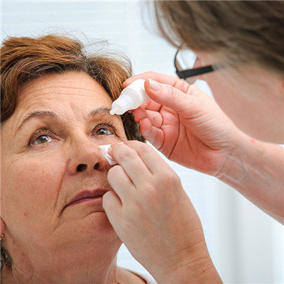
In mild cases, epinephrine is used β Receptor blockers or sympathomimetics may be sufficient; In many severe cases, mydriasis drugs can be added. If the local drug treatment is intolerable or unable to control the intraocular pressure, oral carbonic anhydrase inhibitors can be used.