How is lung cancer left lobar atelectasis to return a responsibility?
summary
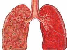
Left atelectasis of lung cancer refers to the decrease of volume or air content of one or more lung segments or lobes. Due to the absorption of gas in the alveoli, atelectasis is usually accompanied by the decrease of light transmittance in the affected area, and the adjacent structures (bronchi, pulmonary vessels and pulmonary interstitium) gather to the atelectasis area, sometimes alveolar consolidation and compensatory emphysema of other lung tissues can be seen. The collateral gas traffic between the lobular segments (sometimes the lobes) can make the completely blocked area still have a certain degree of light transmission. Atelectasis can be divided into congenital or acquired. Congenital atelectasis refers to the fact that there is no gas filling in the alveoli of infants at birth. Clinically, infants have severe dyspnea and cyanosis, and most of them die of severe hypoxia after birth. How is lung cancer left lobar atelectasis to return a responsibility? I'd like to share my views with you.
How is lung cancer left lobar atelectasis to return a responsibility?
Dyspnea cyanosis, and even blood pressure, tachycardia, fever, occasionally can cause shock. Slowly formed atelectasis can be asymptomatic or mild. Middle lobe syndrome is usually asymptomatic, but it often has severe irritative dry cough. Some clinical conditions may indicate the possibility of bronchial obstruction and atelectasis. If some asthmatic children have persistent wheezing, atelectasis may occur. If there is fever at this time, it is suggested that the diagnosis of allergic aspergillosis with mucus impaction is mainly found in asthmatic patients. Fever and tachycardia (pneumonia after surgery) occur 48 hours after surgery, which is often caused by atelectasis. Left lower lobe atelectasis is most likely to occur after cardiac surgery. The patients with chest wall disease can not cough effectively, which is the risk factor of atelectasis. Once the patients have respiratory symptoms, the possibility of atelectasis should be considered. Single or multiple rib fractures can cause atelectasis, especially when there is chronic bronchitis. Children with respiratory symptoms should think of the possibility of foreign body inhalation, especially in the history of choking, choking or coughing. Patients are often unable to provide such information on their own initiative and need to be excluded by purposeful inquiry. It should be noted that there is a long and short asymptomatic period after foreign body inhalation. Adults can often provide a clear history of foreign body aspiration, except for those who are insensitive or delirious.
2. Atelectasis in bronchogenic carcinoma is mainly seen in middle-aged or elderly men with a history of smoking, often with a history of chronic cough. This kind of situation often accompanied by infection, patients often have fever, chills, chest pain and expectoration, repeated a small amount of hemoptysis, more characteristic tumor metastasis to the chest can appear obvious symptoms. The age of onset of bronchial adenoma was younger than that of lung cancer. The respiratory symptoms were nonspecific, but most of them had hemoptysis. Occasionally, patients may present with carcinoid syndrome, suggesting extensive metastasis of the tumor. If the history of pulmonary tuberculosis, pulmonary fungal infection, foreign body inhalation or chronic asthma, we should pay attention to whether there is bronchial stenosis. If you have a history of chest trauma before, you should pay attention to exclude the undetected bronchial laceration and stenosis. About 50% of patients with atelectasis secondary to broncholithiasis have a history of coughing up calcified substances. Patients often do not pay attention to them and need doctors' advice. Some patients think that doctors do not believe that they will cough up "stones", so they deliberately omit this history. Other common symptoms of broncholithiasis include chronic cough, wheezing, repeated hemoptysis and repeated pulmonary infection. In addition, patients in ICU are prone to atelectasis.
3. Signs typical signs of obstructive atelectasis include evidence of reduced lung volume (decreased tactile speech tremor, diaphragmatic elevation, mediastinal displacement), percussion turbidity, voice tremor, and decreased or disappeared breath sounds. If a small amount of gas enters the collapsed area, moist rales can be heard. May have the obvious cyanosis and the dyspnea, after the surgery patient, has the characteristic is repeatedly carries the phlegm sound and the weak cough. If the affected area is small or the surrounding lung tissue is fully and effectively compensated for hyperinflation, the signs of atelectasis may be atypical or absent. In non obstructive atelectasis, the main bronchus is still unobstructed, so the voice tremor is often enhanced and the breath sound exists. The upper lobe atelectasis is close to the trachea, and the bronchial breath sound can be heard at the apex of the lung. The signs of lower lobe atelectasis were similar to those of pleural effusion and unilateral diaphragm elevation. The signs related to the underlying diseases found in physical examination can provide diagnostic clues. Pulmonary atelectasis caused by mucus embolism, mucus impaction or bronchial stenosis secondary to asthma, and characteristic expiratory wheezing can be heard by auscultation. Bronchial lung cancer may have clubbing fingers or other signs of metastasis. Atelectasis caused by lymphoma can be found in different parts of the lymph node enlargement. Atelectasis with jugular vein dilatation or distention and liver enlargement often indicate fibrosing mediastinitis. Cardiac murmur, galloping cyanosis or heart failure can be found in compression atelectasis caused by cardiovascular disease. It is easy to find one or more rib fractures in chest trauma by palpation. Flail chest appears when inspiratory. Atelectasis caused by weakness of chest wall muscle often has evidence of basic neuromuscular diseases.
matters needing attention
1. Congenital atelectasis refers to the fact that there is no gas filling in the alveoli of infants at birth, and there are severe dyspnea and cyanosis clinically. Most of the infants die of severe hypoxia after birth. 2. The prognosis of acquired atelectasis, such as cancer and complications, is poor.