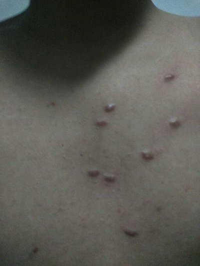
Symptoms of shoulder bursitis
summary
The bursa is a bursa with synovium. Its function is to act as a cushion between muscle cavity and muscle, between muscle cavity and bone, or between skin and bone. There are many types of bursa around the shoulder joint. These bursas are often subjected to chronic stimulation or injury such as friction and impact, which may cause synovial inflammation, hyperemia, edema and exudation of the synovial membrane, swelling and tension of the bursa, resulting in severe pain, and even adhesion of surrounding tissues, adhesion in the bursa, fibrous atresia or calcium deposition.
Symptoms of shoulder bursitis
Acromioclavicular bursa, also known as deltoid bursa, is the most common shoulder bursitis. The acromioclavicular bursa is located below the acromion and acromion ligament, above the rotator cuff and rib tubercle. The top of bursa is attached below the acromion and acromion ligament, and the bottom is attached above the rib tubercle and rotator cuff. In most cases, subacromial bursitis is secondary to adjacent tissue lesions, especially after supraspinatus myositis or supraspinatus leg rupture.
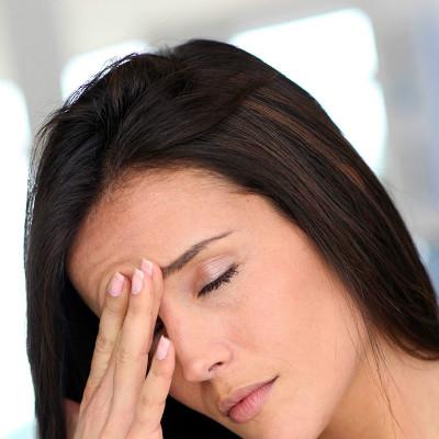
Pain, limited movement and limited tenderness are the main symptoms of subacromial bursitis. The pain increased gradually, especially at night. Especially when the shoulder is abducted and rotated, the pain is aggravated. It is usually located in the depth of the shoulder and involves the insertion of deltoid muscle. It can also radiate to the shoulder, neck, hand, etc. Tenderness points are mostly located in the shoulder joint, subacromial, greater tubercle, etc., which can often shift with the rotation of the bone. When the bursa swells or accumulates fluid, there is tenderness in the area of shoulder joint or deltoid muscle. In order to relieve pain, patients often make the shoulder in adduction and internal position. With the thickening and adhesion of the wall of the bursa, the range of motion of the shoulder joint gradually reduced to complete disappearance, and atrophy of the shoulder 5p band muscle could be seen in the late stage.
Primary subacromial bursitis can be secondary to acute injury or cumulative injury, such as long-term arm lifting labor to make the acromion slide down, and form traumatic bursitis by repeated friction and compression between the acromion and the big tubercle. Most of the pathological changes were inflammation and edema of the bursa, resulting in thickening of the bursa wall, vascular hyperplasia, secondary fibrosis of the bursa wall, and sometimes adhesion and closure.
matters needing attention
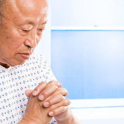
X-ray examination occasionally showed calcification of supraspinatus muscle. Acute injury caused by deltoid bursitis, often a few days after the injury before the symptoms of acute bursitis, acromion bursa puncture according to the amount and character of fluid is helpful to diagnose the nature and degree of lesions.