Oral candidiasis symptoms?
summary
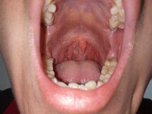
Oral Candida albicans infection is a kind of oral mucosal disease caused by Candida infection. Oral Candida albicans infection is also a common disease in our daily life. For many people, there are many causes of this disease. If we do not pay attention to oral hygiene and abuse antibiotics, oral Candida albicans may be infected, If oral Candida albicans occurs, patients should pay attention to timely treatment to avoid aggravation of the disease. Today we will share the symptoms of oral Candida albicans infection.
Oral candidiasis symptoms?
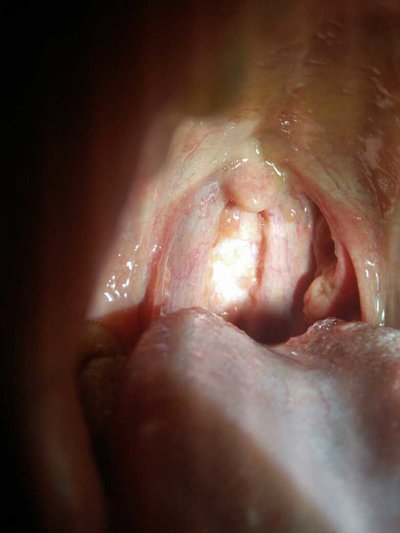
Acute pseudomembranous (snow mouth disease) thrush of newborn usually occurs within 2-8 days after birth. The most common sites are buccal, tongue, soft palate and lip. The mucosa in the damaged area is congested. There are scattered soft spots as white as snow, such as the size of cap needle. Soon they fuse into white or Blue White Velvet patches, and can continue to spread. In serious cases, they affect tonsil, pharynx and gingiva. Early mucosal congestion is more obvious, so it is bright red and white contrast. The old lesions were hyperemia, white patches with light yellow. Patch attachment is not very close, a little force can be wiped off, exposed red mucosal erosion surface and mild bleeding. The children were restless, crying, difficult to lactate, sometimes had mild fever, and the general reaction was mild; However, in a few cases, it may spread to the esophagus and bronchus, causing candidal esophagitis or pulmonary candidiasis. A few patients can also be complicated with children's generalized cutaneous candidiasis and chronic mucocutaneous candidiasis.
Acute erythematous adult acute candidal stomatitis may have pseudomembrane and angular stomatitis, but sometimes the main manifestations are mucosal hyperemia and erosion, mass atrophy of tongue dorsal papilla and thickening of surrounding tongue coating. Patients often first have abnormal taste or loss of taste, dry mouth, mucous membrane burning pain.
Chronic hypertrophic type of buccal mucosa lesions, often symmetrically located in the triangle area of the medial corner of the mouth, nodular or granular hyperplasia, or fixed tight white keratin plaque, similar to the general mucosal leukoplakia. Palatal lesions may develop from denture stomatitis, with papillary hyperplasia of mucosa; The lesions on the back of the tongue can be characterized by filamentous papillary proliferation and gray black color, which is called hairy tongue, so hairy tongue also belongs to this type.
matters needing attention
Remove the food fillings between teeth in time. In the process of chewing food, food fibers are often sandwiched between teeth, which are harmful to teeth and periodontal tissue. We can brush our teeth to keep our mouth clean.